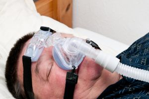
According to a study published online in Neurology®, the medical journal of the American Academy of Neurology, obstructive sleep apnea, a condition that causes low oxygen levels during sleep, is associated with the degeneration of brain regions related to memory. The study found that the changes in the brain were strongly related to the extent of oxygen decline during REM (rapid eye movement) sleep. The study does not prove that sleep apnea causes this degeneration; it merely shows an association.
Obstructive Sleep Apnea is a Sleep Disorder
Obstructive sleep apnea occurs when the muscles of the larynx relax during sleep and block the airway, causing the person to wake up repeatedly to breathe. This disturbed sleep pattern can lower oxygen levels, which in turn can damage small blood vessels in the brain. REM sleep is the stage during which most dreams occur and is associated with numerous critical functions during sleep, including memory consolidation and the processing of emotional experiences.
“Obstructive sleep apnea is a sleep disorder that increases with age, and low oxygen levels during sleep can impair the ability of our brain and body to function properly,” said study author Bryce A. Mander, PhD, of the University of California Irvine. “Our study found that low oxygen levels in obstructive sleep apnea, especially during REM sleep, may be associated with cognitive decline because the small blood vessels in the brain are damaged and this damage affects parts of the brain associated with memory.
The study involved 37 people with an average age of 73 who had no cognitive impairments. They did not take any sleep medications. Sleep studies were conducted on the participants overnight. Of the group, 24 people had obstructive sleep apnea. The researchers measured the participants’ oxygen levels throughout the night in all stages of sleep, including REM sleep. Brain scans were performed on the participants to measure brain structure.
Link to Aging and Alzheimer’s Disease
The researchers found that lower oxygen levels during REM sleep were associated with higher levels of white matter hyperintensities in the brain. White matter hyperintensities are bright spots that appear on brain scans and are thought to indicate damaged white matter tissue. This damage can be caused by injury to small blood vessels in the brain. The minimum oxygen saturation of the blood during sleep and the total time during which the blood oxygen level is below 90% allow conclusions to be drawn about the total amount of white matter hyperintensities in the brain. A blood oxygen level of 90% or less is cause for concern.
The researchers also measured the volume of the hippocampus and the thickness of the entorhinal cortex, both areas associated with memory. They found that more white matter hyperintensities were associated with reduced volume and thickness in these areas. Participants took a memory test before and after sleep to assess sleep-dependent memory. The researchers found that deficits in sleep-dependent memory were associated with reduced thickness of the entorhinal cortex. “Taken together, our findings may partially explain how obstructive sleep apnea contributes to cognitive decline associated with aging and Alzheimer’s disease through the degeneration of brain regions that support memory consolidation during sleep,” Mander said. One limitation was that the study participants were mainly white and Asian, so the results may not apply to other populations.


