During REM sleep, the brain exhibits an unusual combination of phenomena: Blood flow increases, and levels of an important metabolic component also rise—while at the same time, ATP levels—the brain’s primary energy source—decrease in nerve cells. Researchers at Tohoku University have studied this relationship in sleeping mice in real time. The results show that the brain begins preparing for this unique phase even before REM sleep begins.
The Brain Must Operate on a Limited Energy Budget
Sleep by no means means that the brain stops working. On the contrary: During certain sleep phases, the activity of the nervous system changes fundamentally. REM sleep is particularly striking; it is characterized by rapid eye movements and is often associated with especially vivid dreams.
This sleep phase is therefore also referred to as “paradoxical sleep.” The body is largely in a state of muscular rigidity, while brain activity in many respects is more reminiscent of the waking state. This raises an interesting question for researchers: How does the brain supply its neurons with energy when activity changes so drastically within just a few minutes? A new study in Communications Biology provides some surprising insights into this. The researchers observed in mice that the brain’s energy supply changes even before the transition to REM sleep. At the same time, the actual energy consumption of the neurons appears to follow a different pattern.
 Both the human and animal brains are among the body’s most energy-intensive organs. Billions of neurons must constantly generate electrical signals, transmit information, and maintain their connections with one another. These processes require energy. An important energy carrier is ATP, short for adenosine triphosphate. Neurons need ATP, among other things, to restore their ionic balance after electrical signals and to maintain communication between cells.
At the same time, the brain has only a limited amount of energy at its disposal. Therefore, energy supply must be closely tailored to the current activity. It is precisely this adaptation that particularly interests the researchers: Does the brain adjust its energy supply depending on its current state? The transition between different sleep stages offers a particularly suitable model for studying this. During sleep, activity, blood flow, and metabolism change in characteristic ways.
A Look Inside the Sleeping Brain
To directly track these changes, the researchers developed a method that allowed them to observe the brains of sleeping mice. To do this, they coated the animals’ skulls with a transparent, UV-curable material. This enabled the scientists to use imaging to track the activity and blood flow in the brain during natural sleep.
Using wide-field fluorescence imaging, they examined, among other things, changes in blood volume in the brain. This provides insights into how the supply to nerve tissue changes during different states of activity. In addition, the researchers examined two metabolic markers. One of these was ATP in the nerve cells. The second was pyruvate in astrocytes.
Astrocytes are non-neuronal cells of the nervous system that are involved, among other things, in supplying and supporting nerve cells. Pyruvate, in turn, is central to important metabolic pathways and is produced, among other things, during the breakdown of glucose. Thus, its concentration can provide insights into how energy metabolism in the brain changes. The combination of these measurements allowed the researchers not only to observe brain activity but also to track various aspects of its energy metabolism simultaneously.
Blood Flow Changes Even Before REM
Of particular interest was the transition from non-REM sleep to REM sleep. During non-REM sleep, characteristic slow brain activity occurs. Among other things, the researchers focused on fluctuations in the theta frequency range. These changes were associated with subsequent changes in blood volume in the brain. Even more striking, however, was what happened immediately before REM sleep.
About 50 seconds before the classically defined onset of the REM phase, blood volume in the brain began to rise. The change initially began in the posterior region of the cortex and then spread further forward. This suggests that the transition to REM sleep does not simply begin with a sudden switch. Rather, the brain appears to prepare for this state in advance. The increased blood flow could help adapt the supply to the active neural tissue to the upcoming demands.
More Pyruvate, But Less ATP
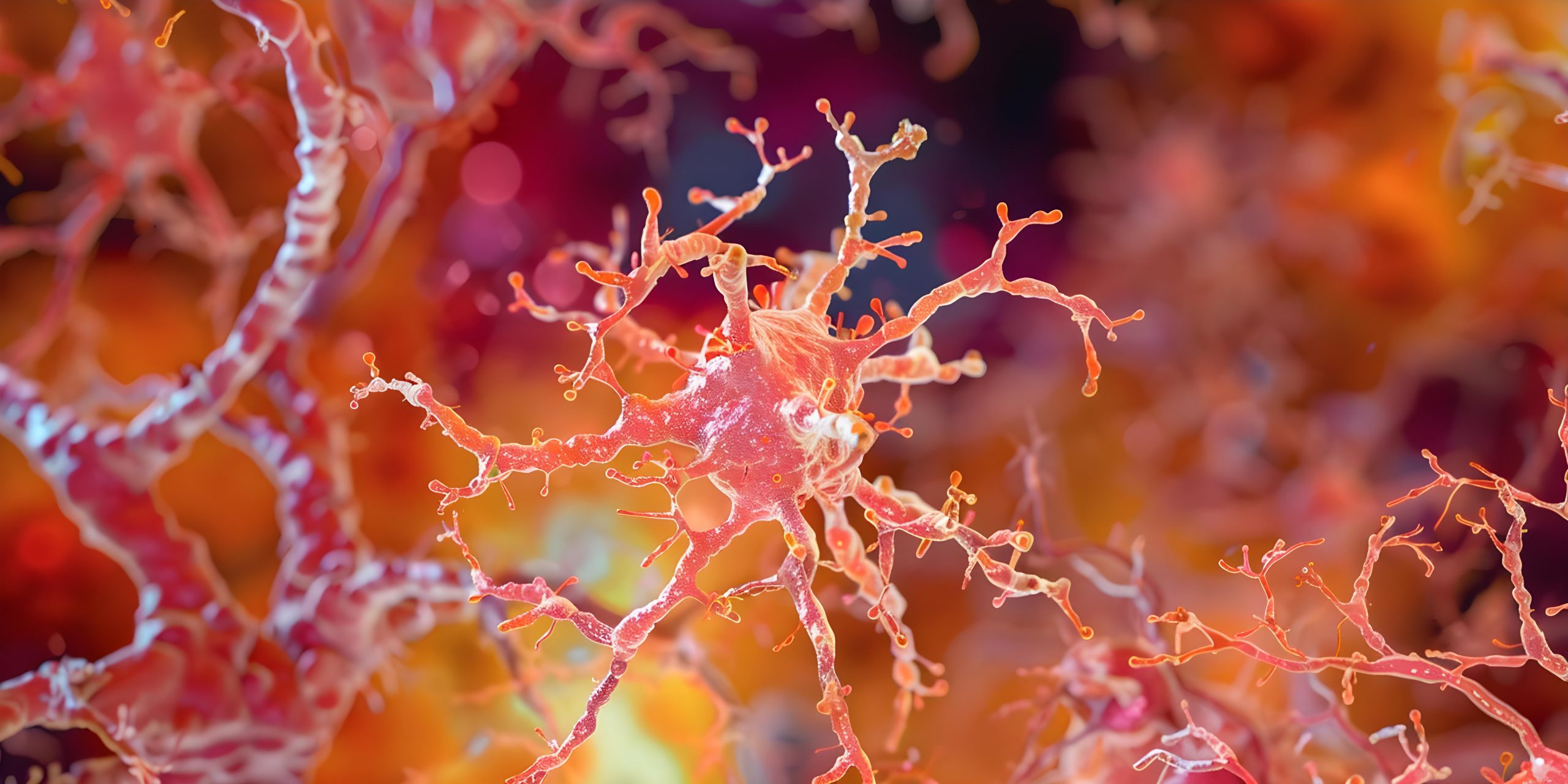
With the onset of REM sleep, a particularly unusual pattern then emerged. The concentration of pyruvate rose in the astrocytes. This could indicate that more metabolic substrate is available during this phase or that the glycolytic activity of these cells increases. At the same time, however, the researchers observed a decrease in ATP levels in the nerve cells.
 At first glance, this seems contradictory. If more blood is flowing through the brain and, at the same time, an important metabolic building block is increasing, one might expect that more immediately available energy would also be present. Yet this was precisely not the case in the mice studied.
Rather, the results suggest that energy supply and energy consumption do not simply run parallel during REM sleep. The brain can increase its energy supply while simultaneously exhibiting lower ATP levels in the neurons.
Do Nerve Cells Consume Particularly Large Amounts of Energy While Dreaming?
One possible explanation is that nerve cells consume a particularly large amount of ATP during REM sleep. REM sleep is associated with complex changes in neuronal activity. At the same time, it is believed to play important roles in memory and information processing. During this phase, therefore, particularly energy-intensive processes could be taking place in the neural networks.
For example, an increased energy requirement for changes at synapses is conceivable. Synapses are the contact points between neurons and play a central role in learning and the storage of information. Communication between different brain regions could also play a role during REM sleep. In particular, the interaction between the hippocampus and the cerebral cortex is being studied in connection with the processing and consolidation of memories.
However, the new findings do not prove that these specific processes are responsible for the observed decline in ATP. The researchers cite several possible mechanisms that need to be examined more closely in future studies. In addition to increased consumption, a change in the transport of substances within the brain is also a possibility. Astrocytes and nerve cells work closely together in energy metabolism. Astrocytes can provide metabolic byproducts that are reused by neurons. If this collaboration changes during REM sleep, it could explain why the increase in pyruvate in astrocytes does not immediately lead to higher ATP levels in the neurons.
Mitochondria could also be involved. These cell organelles produce a large portion of the ATP in the cells. Changes in their activity could likewise influence how much ATP is actually available to the neurons. Based on the current results, it is not yet possible to determine which of these explanations is correct.
The Brain Apparently Begins Preparing Even Before REM
The timing of the observations is particularly interesting from a chronobiological perspective. The increase in blood volume began approximately 50 seconds before the transition to REM sleep. This suggests that the changes do not arise solely as a reaction to the REM state that has already begun.
 Instead, the brain may be actively preparing for an impending change in state. The transition between sleep stages would thus not only be a consequence of altered neural activity but possibly a coordinated process in which the brain’s energy supply also changes proactively.
This is consistent with a fundamental principle of biological timing: Organisms do not react exclusively to changes that have already occurred. Many physiological processes are temporally coordinated and can prepare for an impending state.
Why These Findings Are Interesting for Sleep Research
Sleep is much more than a period of rest. During the night, different states alternate, each with characteristic patterns of neural activity, metabolism, and blood flow. REM sleep occupies a special position among these. Although the muscles are largely at rest, the brain exhibits complex patterns of activity. The new observations add another layer to this picture: the brain’s energy metabolism also changes during the transition into this sleep phase. It becomes clear that “increased energy supply” does not automatically mean that individual neurons have more ATP available. Supply, metabolism, and actual energy consumption can be out of sync. It is precisely these differences that could help us better understand how the brain maintains its functions under changing conditions.
The results come from experiments on mice. They therefore initially show how the metabolic and circulatory processes examined in the mouse brain change during different sleep states. Whether the observed processes occur in the same way in humans remains to be investigated. Furthermore, it is not immediately clear from the measurements what specific role the observed decline in ATP plays in dreaming or memory processing. It also remains unclear whether the changes are the same under different sleep conditions, at different ages, or in other physiological states.
Further studies could therefore clarify how the various cell types cooperate with one another during REM sleep and how the temporal coordination between neuronal activity, blood flow, and metabolism functions. The study thus provides further evidence that the brain regulates its energy balance extremely dynamically during sleep. In particular, the transition to REM sleep appears to be associated with a preparatory change in blood flow and a subsequent reorganization of metabolism.
The apparent paradox—more signs of energy supply while simultaneously having less ATP in the nerve cells—could indicate that the brain during REM sleep relies particularly heavily on a flexible allocation of its resources. This also makes it clear why sleep is by no means a passive pause for brain function. While we sleep, the brain changes its operating state, reorganizes its activity, and adjusts its energy supply. REM sleep demonstrates particularly impressively just how complex this nocturnal regulation actually is.
You may also be interested in...
-

There has been a great deal of buzz in the medical community regarding circadian rhythm…
-
Caffeine is not only found in coffee, but also in tea, chocolate, energy drinks, and…
-
Do sharks nap? Perhaps more importantly, why does it matter? As explained by researcher and…